 Contact
 Intranet
 SK
SK


Phase contrast X-ray imaging of light weight samples
Weakly absorbing objects (such as biological, polymers, etc.) typically have 1000 times stronger contrast in the X-ray region in the so-called phase-contrasted imaging mode as compared to the absorption imaging mode. This was the motivation for the X-ray microscopy setup we developed, which is based on an X-ray microfocus generator attaining a spatial resolution down to 3 μm with an average lateral coherence length of 0.3 to 13 μm and up to a 140-fold geometrical magnification [1,2]. The X-ray imaging system allows phase contrast imaging of light weight samples, e.g. bilogical samples, wood (Fig. 1), plastics (Fig 2a), paper, and so on. However, such a setup uses broad polychromatic radiation, therefore, the extracted quantities from the X-ray images, such as attenuation coefficient and the retrieved phase (Fig 2b), are affected by beam hardening effects and the X-ray images have usually a reduced contrast due to the presence of scattering [3]. The use of monochromatic radiation will, therefore, be of benefit but at the cost of a significant photon flux loss. However, the recent developments of more intense laboratory X-ray sources using liquid anode in combination with collimation optics and high-efficient direct conversion integrating or single photon counting X-ray detectors allow us to employ the X-ray crystal optics for imaging applications in laboratory conditions in an efficient way. Such a setup will allow for quantitative scattering-free X-ray imaging.
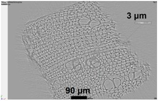
Fig. 1 CT slice with recognizable structural areas of wooden cells with a wall thickness of 3 micrometres.
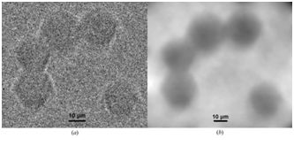
Fig. 2 (a) Single X-ray projection of PMMA spheres (diameter of 20 mm) after flat-field correction and (b) the retrieved phase image (relative electron density) of PMMA spheres.
Zápražný, Z., Korytár, D., Dubecký, F., Áč, V., Stachura, Z., Lekki, J., Bielecki, J., and Mudroň, J.: Experience with imaging by using of microfocus x-ray source, J. Electrical Engn. 61 (2010) 287-290.
Zápražný, Z., Korytár, D., Áč, V., Konopka, P., and Bielecki, J.: Phase contrast imaging of lightweight objects using microfocus X-ray source and high resolution CCD camera. (2012), JINST 7 C03005.
Zápražný, Z., Korytár, D., Mikulík, P., and Áč, V.: Processing of projections containing phase contrast in laboratory micro-computerized tomography imaging. J. Applied Crystall. 46 (2013) 933-938.